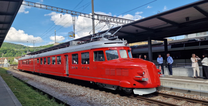
Switzerland’s first SPARC Compact CL system
CL Delmic
18.06.2026
Switzerland’s first SPARC Compact CL system

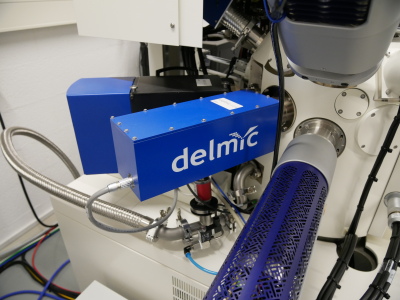
Switzerland’s first SPARC Compact CL system – now in use at the University of Lausanne.
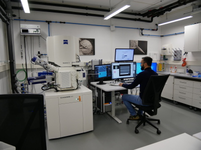
The Faculty of Geosciences and Environment (FGSE) is unique in Switzerland: it is dedicated to researching natural and anthropogenic changes to our planet, particularly in Alpine regions and urban centres. In doing so, it brings together various geoscientific disciplines to understand the processes that shape our environment and landscapes.
The recently installed SPARC Compact cathodoluminescence (CL) system, integrated into a ZEISS Gemini scanning electron microscope, opens up new analytical possibilities, particularly for researchers at the Institute of Earth Sciences. Minerals store information about their origin and development in their chemical composition and crystalline structure.& nbsp;CL analysis makes these properties visible and allows valuable conclusions to be drawn about the formation history of rocks. The data obtained can also make an important contribution to determining the age of minerals.
The first BSE and CL images of quartz, zircon and calcite demonstrate how this works in practice:
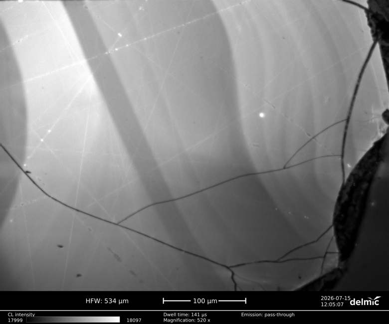
quartz
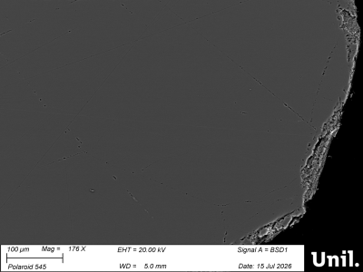
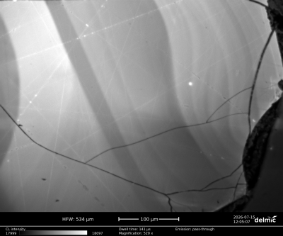
(BSE) of (a) quartz, imaged at an accelerating voltage of 20 kV and a current of 2 nA. (b) Cathodoluminescence (CL) image of a quartz grain, imaged at an accelerating voltage of 20 kV and a current of 1 nA with a dwell time of 140 ms. The CL image shows the growth pattern of the quartz grain.
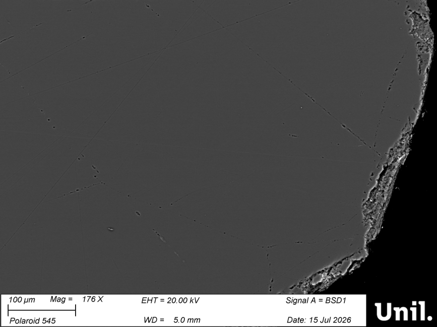
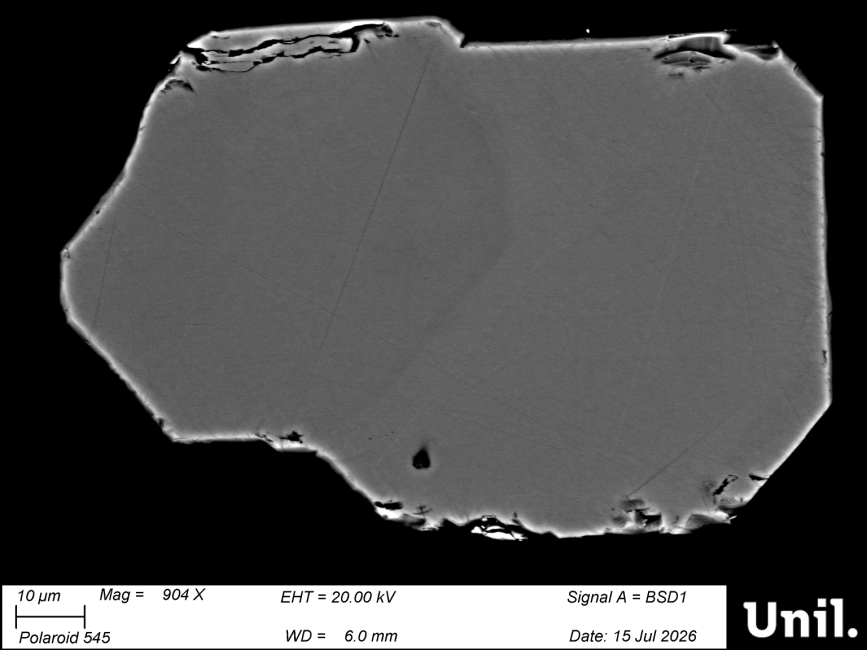
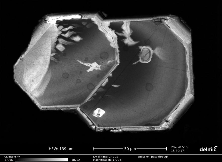
Zircon
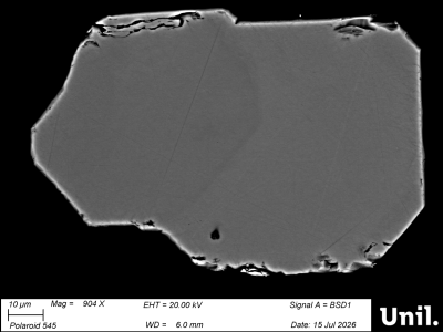
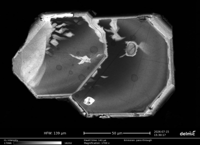
(BSE) of (c) zircon, imaged at an accelerating voltage of 20 kV and a current of 2 nA. (d) CL image of a zircon grain, imaged at an accelerating voltage of 5 kV and a current of 500 pA with a dwell time of 140 ms. The CL image shows a twinned zircon crystal.
Calcite
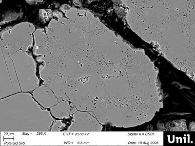
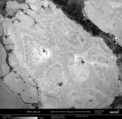
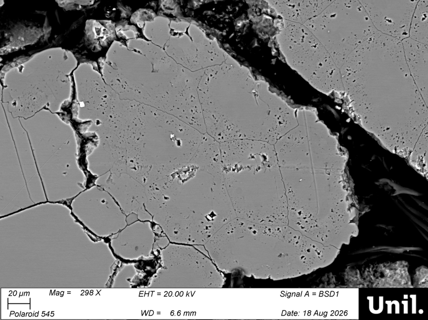
(BSE) image of (e) calcite grains, taken at an accelerating voltage of 20 kV and a current of 2 nA. (f) CL image of a calcite grain, taken at an accelerating voltage of 8 kV and a current of 2 nA, with a dwell time of 400 µs and a 400 nm bandpass filter with a bandwidth of 50 nm. The CL image shows the cell-like microstructures of Microcodium (cells of the root cortex).
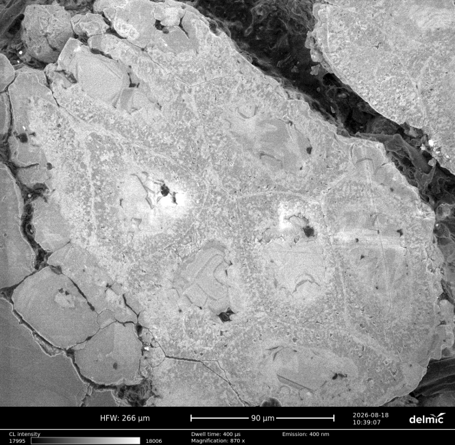
All the images and measurement data shown were provided to us by Dr Goran Andjić.
The images were taken at the Faculty of Geosciences and Environment (FGSE) at the University of Lausanne. The system is integrated into a ZEISS Gemini scanning electron microscope fitted with an ULTIM Max EDS and Symmetry EBSD detector from Oxford Instruments. By combining the SPARC Compact CL system with the ZEISS Gemini SEM, this information can be captured at high spatial resolution and directly linked to the electron microscope images. This opens up new possibilities for investigating the composition and formation of geological materials in even greater detail.
We are delighted to have realised Switzerland’s first SPARC Compact CL installation with this project.
Would you like to find out more about the possibilities of cathodoluminescence, or are you planning a similar project? We would be happy to advise you on the right solutions and options for your application.