RISE

RISE
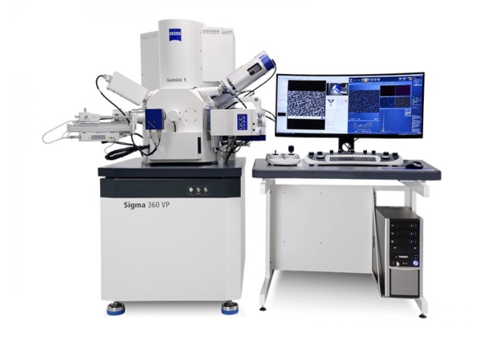
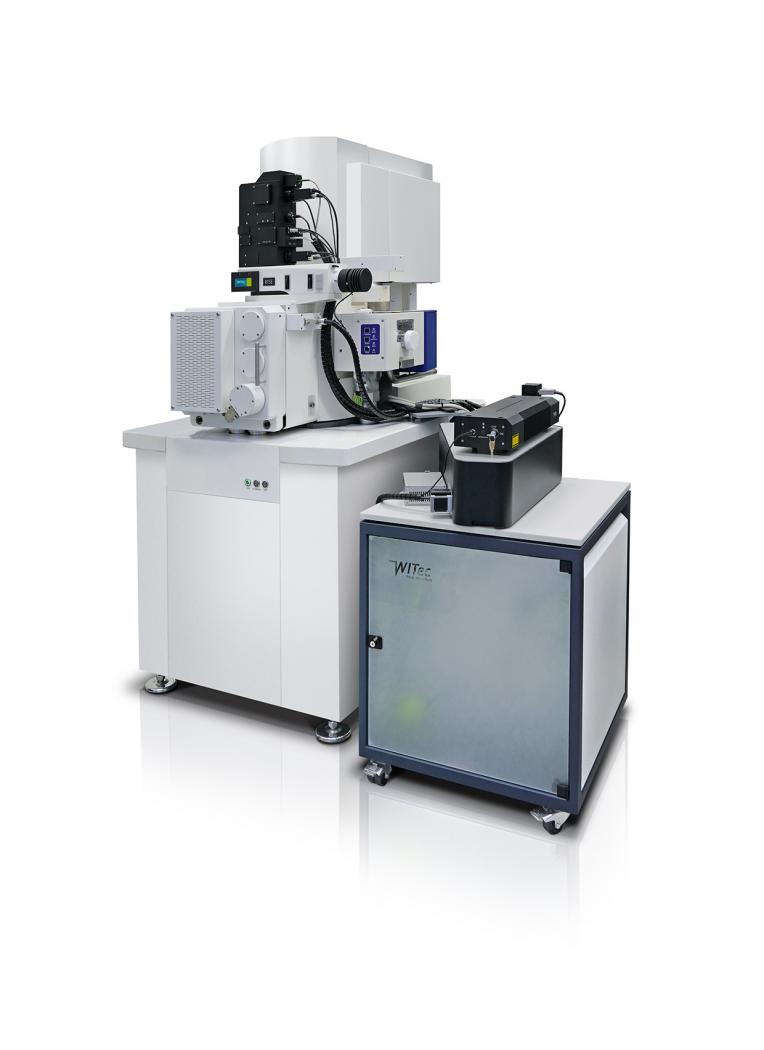
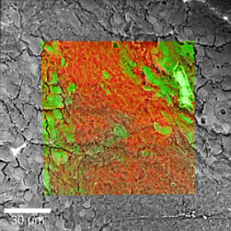
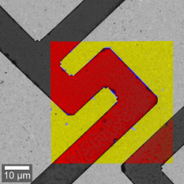
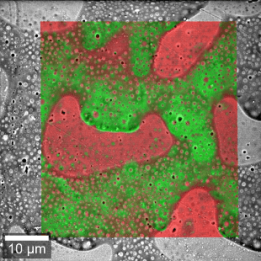
RISE microscopy is a correlative microscopy technique that combines electron-optical methods (Scanning Electron Microscopy, SEM) with Raman imaging. RISE enables information on ultrastructure, surface properties and molecular constituents to be measured at the same sample location, and this data to be correlated.
RISE combines all the features of a stand-alone SEM and a confocal Raman microscope in a single instrument:
- Faster and more user-friendly switching between Raman and SEM measurements;
- Automated sample transfer between the two measurement positions;
- Integrated software interface for user-friendly control of measurements;
- Correlation of measurement results and image superimposition;
- No compromise on SEM or Raman imaging quality.
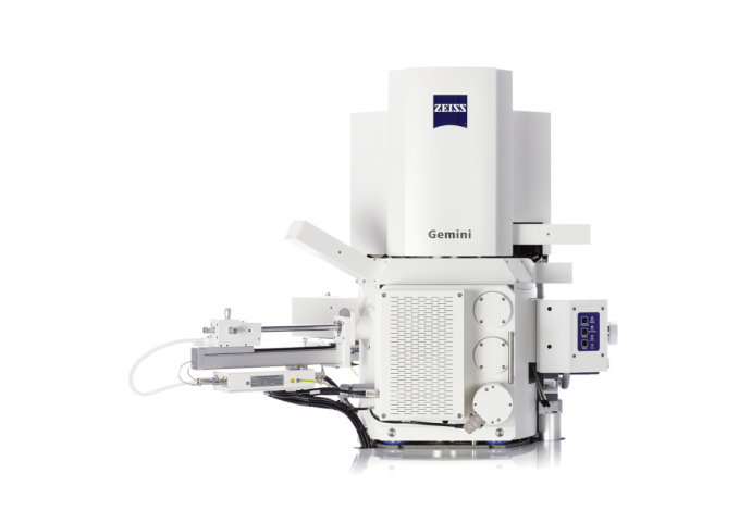
RISE can, for example, be combined with the ZEISS Sigma 360 or the GeminiSEM 460. A Raman system can also be combined with a ZEISS EVO, subject to consultation and subject to space constraints. WITec and ZEISS have jointly developed this system in order to be able to offer a fully integrated instrument as an OEM product via ZEISS. This gives researchers and users even more options for chemical-spectroscopic and structural analysis.
The seamless combination of these two techniques in a single instrument offers a decisive advantage: measurements are quicker and simpler than when using two separate instruments, as the components required for Raman measurements (objectives, stage) are integrated into the SEM’s vacuum chamber. The Raman microscope is connected via a standard port on the SEM. During measurements, the sample is moved from one measurement position to another and remains in the vacuum chamber for the entire experiment. WITec’s professional optical Raman microscope includes a light microscopy function, enabling users to quickly gain an overview of the sample.
The structural and chemical data are analysed and correlated using WITec’s Suite software. This software is intuitive to use and includes automated features for rapid, routine analysis.
Examples of use
We are looking forward to hearing from you