Ultim Max Infinity

Ultim Max Infinity
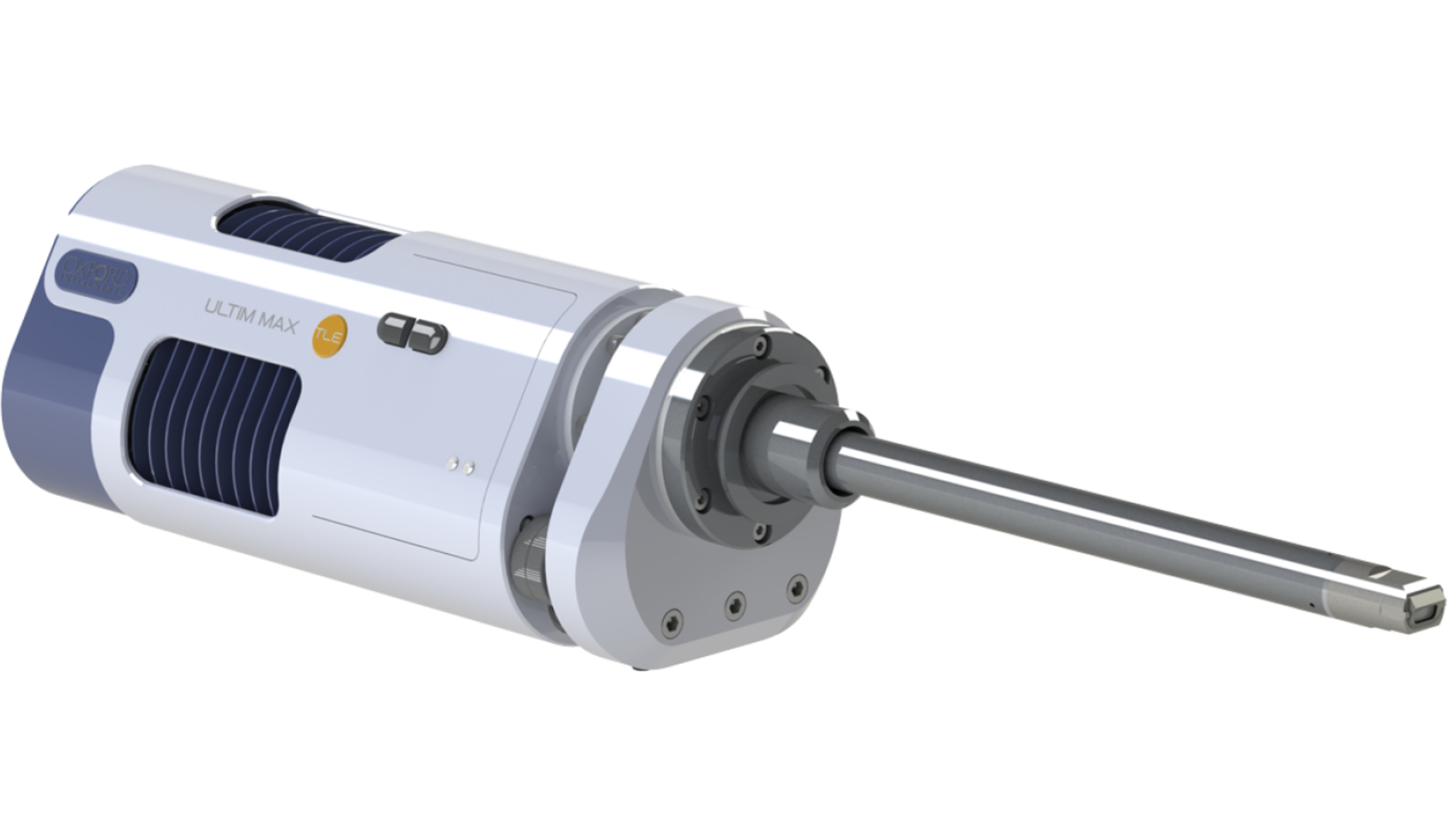
The Ultim Max Infinity EDS detector from Oxford Instruments has been developed for high-resolution chemical element analysis in a scanning electron microscope (SEM). The system combines high sensitivity, fast data acquisition and precise X-ray spectroscopy to reliably determine the chemical composition of materials, particles and nanostructures. It is particularly suitable for applications in materials science, semiconductor research, nanotechnology and industrial quality control.
With sensor areas ranging from 40 to 170 mm², the Ultim Max Infinity captures sufficient X-ray signals for detailed analysis of small structures, even at low acceleration voltages. The high energy resolution enables the reliable differentiation of closely spaced elemental peaks and supports the investigation of light elements, complex material phases and low element concentrations. Tru-Q IQ signal processing optimises spectrum evaluation individually for each detector.
In combination with the AZtecLive software, the Ultim Max Infinity enables real-time chemical imaging as well as quantitative elemental analysis and high-resolution EDS mapping. Chemical distributions can be precisely visualised through the correction of signal overlap and measurement artefacts. The system thus supports both the rapid examination of large sample areas and the detailed characterisation of complex micro- and nanostructures.
- EDS/EDX detector for qualitative and quantitative elemental analysis in SEM
- Sensor areas of 40, 65, 100 and 170 mm²
- Energy resolution of ≤ 46 eV at C Kα and 50,000 cps
- Energy resolution of ≤ 127 eV at Mn Kα and 200,000 cps
- Element detection from beryllium (Be) to californium (Cf)
- Real-time chemical imaging and EDS mapping with AZtecLive
- Individually optimised spectrum processing with Tru-Q IQ
Product overview
Watch the video to find out more about the Ultim Max Infinity and gain an insight into EDS detector technology, high analysis speeds and the possibilities for precise chemical element analysis using a scanning electron microscope SEM.
We are looking forward to hearing from you